The Practice: +44 (0)1638 782000 | The Hospital: +44 (0)1638 782020

Diagnostic Imaging

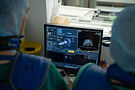
Diagnostic imaging services provide a comprehensive spectrum of capabilities for both first opinion and referral patients. These include radiography (X-ray) and ultrasound using Canon DR and Logiq Fortis capabilities respectively which provide exceptional image quality. The hospital radiography suites have both fixed gantry and mobile generators enabling imaging to be performed from the spine to the foot. Advanced diagnostic imaging techniques provided are nuclear scintigraphy (bone scan), magnetic imaging (MRI) and computed tomography (CT).
Nuclear scintigraphy is a screening imaging technique for the entire skeletal system which uses a radiopharmaceutical (radionuclide and targeting molecule) to identify bone which is undergoing an increased osteoblastic process whether due to fracture, contusion, neoplasia or an adaptive response to exercise or loading. The radiopharmaceutical is injected into the patient’s vein and the targeting molecule guides the radiation emitting radionuclide to the bone. The gamma camera then identifies the radioactivity producing ‘hot spots’ on the generated image. This imaging technique is sensitive for detecting stress fractures in racehorses and is also used for patients with poor performance or elusive lameness. While x-rays detect structural change, if there is an obvious fracture or following significant changes in bone composition or remodelling, nuclear scintigraphy identifies changes at a cellular level which can precede identifiable structural changes.
The onsite system is for a standing sedated patient and enables evaluation of structures between the foot and carpus (knee) or tarsus (hock). It does not involve ionising radiation. MRI uses magnetic fields and radio waves to measure how much water there is in the different tissues of the imaged area. The differing sequences and planes used map a detailed picture of both bone and the soft tissues which includes ligaments, tendons, fascia and joint capsules. The process of acquiring images can be lengthy (2-3 hours) depending on the area to be imaged and the patient’s ability to stand still and square. It is therefore important that the area needing to be assessed has been carefully localised in advance. This is a targeted imaging process not a screening tool. It identifies injuries but also can detect early changes in bone and soft tissues that can aid prevention of lesion progression.
Computed tomography is a form of 3 dimensional X-ray where multiple very thin slices of tissue information are rapidly acquired and following computer processing a detailed high resolution 2D image or 3D reconstruction is produced. NEH has exceptional CT capabilities by having both a fixed large bore Qalibra machine and a mobile in theatre Ceretom. The Qalibra enables rapid image acquisition in the standing patient of the head, lower limbs and upper two thirds of the neck. Under general anaesthesia the large bore of the machine allows increased body coverage and can be used for detailed surveys of the spine and limbs. The capabilities also extend to CT guided surgery. The mobile in theatre Ceretom facilitates pre-operative assessment and surgical planning together with intra-operative guidance and post-operative imaging. Having access to both machines affords excellent surgical support optimising decision making, accuracy and guidance.

Have an emergency?
If your horse needs urgent veterinary attention, our team is ready to help around the clock. Call 01638 782020 for the hospital and 01638 782000 for the practice.